>>> Click here to access this episode of the Syllab Podcast on Spotify <<<
a) How to think of body organs and systems
As we debut this second series, it is important to highlight a few things. The first is that many concepts developed during the Series 1 will be put to good use once again. Sometimes I will provide a brief reminder of the essence of such concepts but not always, thus if you have not previously read or listened to the 10 chapters it comprises, I would strongly advise you to do so. Secondly, Series 1 also contained crucial sections about the notion of function, information (see S1 Section 3.a) and evolution (see S1 Section 3.e) that provide an intellectual framework about the fundamental dynamics of biological organisms. Thirdly, we already followed the complexity build-up all the way from elementary particles up to the cell and even quickly discussed the concepts of cell differentiation and coordination within organisms and the fitness advantages thus provided when confronted with specific environments. Indeed, natural selection is a real thing and eventually most mutations do not make the cut into posterity.
Hopefully, the coming sections and chapters will convey in adequate and novel fashion how cool biology is, not words I would have uttered coming out of high school.
This preamble is applicable not only for this first chapter but for the entire Series 2 as we consider the various systems, organs and senses of the human body, that which marks the formal limits of our biological organisms. Perhaps the easiest way to transition from the previous series into this new one is to define the terms of organs and biological systems, or rather to supply the mental template for how to think about them.
An organ is effectively the hardware of an organism characterized by a defined location and by marked cell differentiation that enables this particular set of cells, with dimensions that vary tremendously, to implement specific functions with high efficiency. Something an undifferentiated cell or even a network of them would be unable to accomplish. However, this should not be understood to mean that one organ performs only one function but rather that organs have evolved in a manner which makes them excel at one or several functions. Ultimately, an organ is still an abstract concept of the human mind so it can easily be subdivided into regions performing different tasks, and it is effectively what happens when you start diving into technical books and papers.
The use of the term “hardware” in relation with organs was a bit of a giveaway and yes, biological systems, which we can crudely divide into organ systems and sensory systems, can and should be thought of as “software” ensuring multiple tasks are carried out in a coordinated and complementary fashion. As such, they not only involve organs but also communication structures, signalling mechanisms, and information processing and dispatching centres.
Just like there are to this day discussions around whether specific parts of the body form an organ or not, the list of systems present in the human bodies is not set in stone, nonetheless there is a strong consensus regarding the delineation of organ systems into 11 major networks and for sensory systems the number is five, or six, or seven, or eight depending on whom you ask. I belong to team 8 senses and so will present them accordingly.
- This Chapter 1 will focus on the respiratory and circulatory systems including their role in gas exchange through breathing, the capillary network, and the role of blood in transporting important molecules across the body and helping achieve homeostasis (refer to S1 Section 5.e).
- In Chapter 2, we will turn our attention towards the digestive system, the solution devised by evolution to transform the molecular makeup of the energy contained in external nutrients so that this fuel can be used in various parts of our organism. We will then enquire into the renal or urinary system involved in the excretion of fluids and the overseeing of blood volume and pressure. It’s all interconnected.
- In Chapter 3, we’ll direct the spotlight onto the musculoskeletal system which integrates the skeleton and the muscles and makes movement, the macroscopic translation of our body in space, possible. Since we’ll be dealing with organic tissues, this will be a good time to also discuss the skin, the wrap-around tissue forming the largest organ in our anatomy.
- Completely different topics for Chapter 4 during which we will first revert to the matter of hormones and their signalling role as part of the endocrine system before studying the reproductive system but, since we have already covered the key aspect of the advantages of sexual reproduction from an evolutionary standpoint (S1 Section 3.e) and the initial phase of embryogenesis post fertilization (S1 Section 3.d), we will be able to concentrate on the organs rather than having to start from scratch and explain some of the foundational ideas.
- Chapter 5 will centre on the immune system whose role is to ensure the detection of pathogens and elicit an internal response such as the set of processes involved when we are fighting off diseases.
- Chapter 6 will be pivotal, literally and metaphorically, and fully devoted to the nervous system, traditionally divided between the central part crowned by the brain and the nerves and ganglia making up the peripheral nervous system. The role of the nervous system is to transmit information, inbound from our senses and outbound to our muscles and other parts of our body, and of course to process this information by interpreting it and making decisions, whether consciously or unconsciously.
- Finally, the last four chapters of the series will be dedicated to the sensory systems whose job it is to gather data primarily about the external world (from the perspective of the organism enclosed within a body), so that it can be interpreted by the central nervous system and acted upon. The exception is interoception which scans the inner state of our body and is thus inward looking. Chapter 7 will be dedicated to vision, Chapter 8 to olfaction and taste, Chapter 9 to audition and touch, and Chapter 10 to interoception and the vestibular sense, the system involved in us maintaining our balance and orienting ourselves.
b) Breathing and anatomy of the respiratory tract
Everything evolves for a reason. This statement should not be understood to mean there is a conscious act behind evolution but, rather, that the chain of mutations leading to the development of a particular genetic trait needs to clear certain hurdles, namely that the new function or improvement in an existing one resulting from these mutations, should have a pay-off in terms of survival and reproduction rate that is higher than the differential in energetic cost associated to this new trait. In other words, the reason behind evolution is that it’s worth it from the perspective of the organism with said mutation.
When we consider the importance of chemical energy, and in particular of the ATP macromolecule described in S1 Section 4.c, it is fairly obvious the more ATP can be produced during a given amount of time, the more work can be done by the organism, which powers not just vital functions but also movement such as the hunting of a prey or the running away from a predator. However, it is not simply about producing more, it is also about doing it efficiently and, in this respect, one of the workhorses of ATP creation is oxygen (O2), primarily on the back of its role as electron acceptor in the electron transport chain (something we saw in S1 Section 7.f on photosynthesis).
So, where is the best place to get oxygen from? Well, it happens to constitute about 21% of the atmosphere so really, it’s there for the taking. Sounds simple, but organisms are made up of cells inside which the ATP is produced and these cells are contained within lipid membranes (refer to S1 Section 4.d) whose very purpose is to separate the inside of the cells from the outside to allow both for internal structure and control over the concentration of certain atoms, ions and molecules, also taking advantage of electrochemical gradients to do work – as a reminder, a gradient is a difference in concentration between two locations and nature’s propensity to equalize this unbalance can be harnessed.
This being the case, there are two challenges to overcome: the first is to get oxygen from the atmosphere and all the way to the edge of the cells, this is called breathing, and the second is to get the O2 molecules across the membrane, this is termed “gas exchange”. Let’s tackle those in this order.
Breathing is undoubtedly the easiest function to describe though arguably not the most straightforward to develop given the interlocking mechanisms and equipment required in terms of muscles, various anatomical elements and regulation. Purely describing the lay of the land, or the lungs in this case, is very top-down and not the best way to help us understand why the human body developed in its seemingly quirky ways. We need to keep our eyes on the concept of functions and efficiency. With that frame of mind, what we would expect to see is a way to bring air inside the body, maximise the interface between that fresh air and either the cells or an intermediary transport medium, and perhaps even use that same infrastructure to evacuate some waste by-products outside of the body. In all honesty, even without hindsight, I think that one could be deduced quite easily. And of course this is what happens.
The diaphragm is a large skeletal muscle located just below the lungs in our thorax, when it contracts, it moves downward, thus expanding the volume of our chest cavity, effectively pumping air in our lungs. This diaphragm movement is supplemented by a contraction of the muscles between our ribs expanding the rib cage upward and outward. Since those muscular tissues are elastic, their subsequent relaxation produces a recoil of the lungs and thoracic cavity which can be enhanced by the activation of another set of muscles (the internal intercoastal and abdominal muscles) during a mechanism called forced exhalation. This is how the air gets in, then out.
In terms of travel, the fresh air goes through the nose and mouth into the trachea and down into the right and left lungs. The lungs are nothing like an empty bag and consist of airways branching into ever smaller conducts terminating into tiny areas where gas exchange takes place. The larger passages are called bronchi, after many divisions they become bronchioles, at which point they cease to be supported by cartilage, and the pulmonary gas exchange occurs inside the alveoli. It’s tricky to visualize but there are close to 500 million alveoli in a human pair of lungs with a total surface area of nearly 80m2 – that’s a rectangle or 8m by 10m or the floor space of a mid-sized apartment.
c) Gas exchange and transport
Here we must highlight that the alveoli do not permeate our entire body so there needs to be an intermediary step between the gas within the lungs and its delivery into the cells. This is the role of the circulatory system and the blood in particular, a topic we’ll move to afterwards. This means the oxygen must get across the blood-air barrier, be transported across the entire body and then cross into the cells where it will be used in the process of ATP generation.
The key concept in understanding how the crossing of membranes by gases can occur is molecular diffusion. It can be explained as the ongoing equalization of concentration between two separate areas, including when separated by a thin membrane. This dynamic has to do with minimization of potential energy but I won’t cover the same ground as in Series 1 so what matters is that the content with high concentration passively flows towards the area with lower concentration, no energy needs to be expanded for this to take place. The main factors in the rate of diffusion include the membrane permeability and the relative gas pressure on each side of the barrier. It should be noted that permeability is selective and is a function of thickness (in the order of 0.0022mm for the blood-air barrier) and of the solubility of lipophilic gases (the organic membranes tend to be lipid bilayers, as explained in S1 Section 4.d); in addition there may also be pores, or protein gas channels, involved in letting gas molecules through. The diagram included below (Figure 1) illustrates the gas exchange between a pulmonary alveolus and a pulmonary capillary, part of the circulatory system.
Figure 1: Gas exchange between alveoli and capillaries
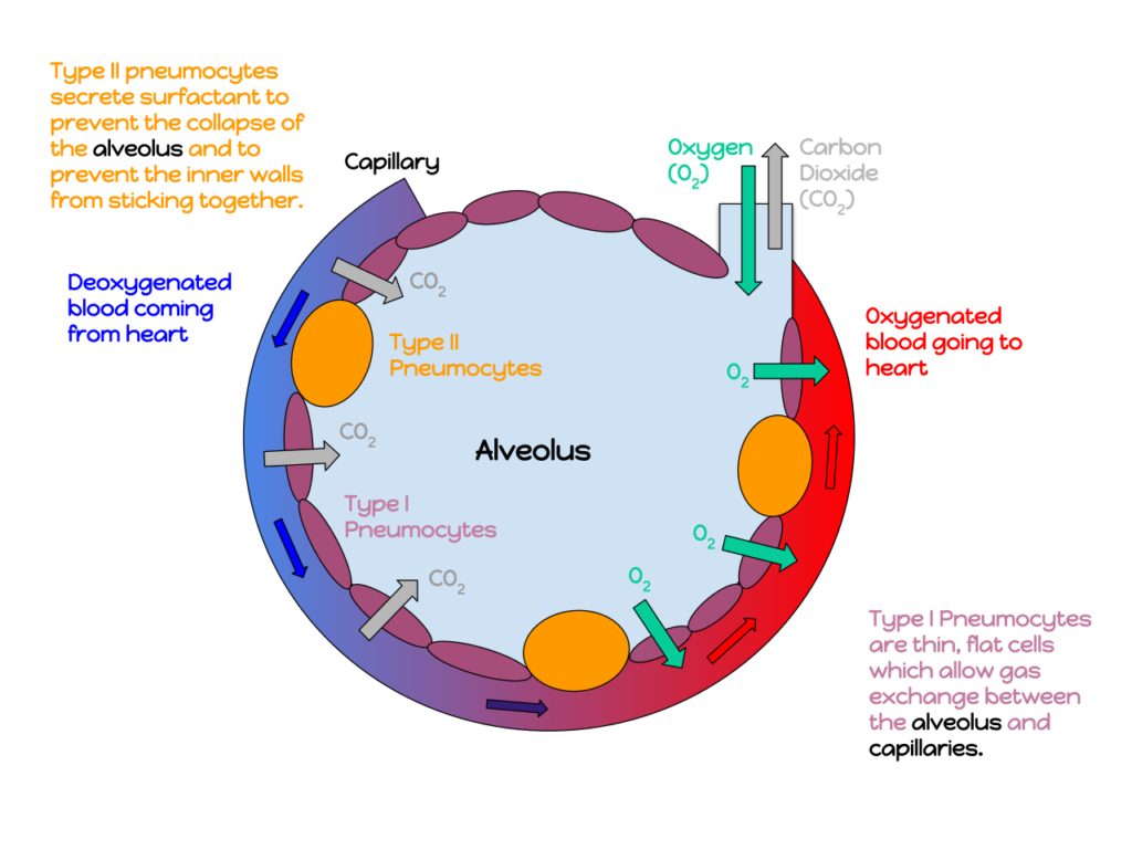
Credit: by Katherinebutler1331 (CC BY-SA 4.0)
Gas pressure requires a little more explanation because it not only drives the diffusion process but it also determines the quantum of exchange taking place. The pressure of a gas is the force applied perpendicular to a specific surface and is a function of the amount of substance it contains, its temperature and it is also inversely proportional to volume. Another way to phrase this is that gas pressure is a function of molecular density and temperature with the latter essentially representing the kinetic energy stemming from the motion of those molecules.
A fluid such as the atmosphere comprises several types of gases and, as physics would have it, it is possible to compute the total pressure by adding the individual pressure exerted by each gas and diffusion will work towards the equalization of each type of molecular concentrations. The important corollary is that the diffusion of both oxygen and carbon dioxide across the blood-air barrier can serve to top up or down the level actually required by the organism to meet its energetic needs and homeostasis.
The absolute value of the following numbers is not important for the sake of understanding but the magnitude of the exchange is interesting. When the blood reaches the alveoli, the oxygen pressure is approximately 6.0kPa and after it leaves it has equalized with the level of the alveoli at 13-14kpa. For CO2, the number decreases from 6.0 to about 5.3, indicating that carbon dioxide is leaving the organism. When there is extra demand on O2, the heart will pump more blood and more oxygen will make its way into the circulatory system and ultimately to the tissue signalling for it. What you may have missed here is that since oxygen accounts for nearly 21% of the atmospheric pressure (as a reminder, 1 atmosphere is 101.3kpa), its ambient pressure when being breathed in is circa 21kpa, not the 13-14kpa of the alveoli. This difference is due to the cul-de-sac nature of the respiratory tract which results in some volume of air called “dead space air” left over after exhalation. This partial rather than full renewal ensures the oxygen pressure does not change dramatically between each breath and in turn makes the control of the inner pressure much easier to achieve. The volume remaining in our lungs after standard exhalation is called functional residual capacity and after forceful exhalation it is called residual volume. If one day you take up free diving, you will recognize those terms.
Water molecules are polar, they have an uneven distribution of electric charges, whereas oxygen is non-polar. Consequently oxygen has low solubility and its transport is facilitated by binding to iron ions (Fe2+) within a protein called haemoglobin, which oxygen rides until delivery to the location in need of it. Once there, the process of diffusion once again kicks in and since the tissues with oxygen deficit have lower oxygen pressure then these molecules will cross the blood vessel membranes, only this time on their way out.
d) The heart and blood vessels
I have mentioned the circulatory system a few times already and now is the right time to look into it a bit more carefully. Functionally, the role of this system is to ensure the circulation of blood within the entire organism where blood takes on the role of transport medium for nutrients and gases towards the tissues, shipping back some of the gases that will be excreted via the lungs, including but not limited to carbon dioxide.
The network of pipes through which blood transits is called blood vessels and, because there are no muscles to force the flow of blood, the circulation relies on pressure provided by the heart whose job it is to pump the blood throughout the blood vessels. I will say very little else about the heart but have included a link to the Wikipedia entry on cardiac cycle at the end of the chapter for any reader interested to learn more.
Unsurprisingly, given what we have already observed in the case of the lungs and the reliance on the physical process of diffusion, the blood vessels start as rather large tubes called arteries and bifurcate into arterioles then keep branching into capillaries with extremely thin membranes, like those in contact with the pulmonary alveoli. This is where the exchange of gas, nutriment and metabolic waste takes place, between the blood and the cells. Unlike in the lungs where the alveoli are dead end spaces, the blood flows through the capillaries so the arterial part of the circulatory system has a symmetrical alter-ego: small venules connect to the capillaries and merge into larger veins that make their way back to the heart.
The artery-out and vein-back dichotomy is almost matched by oxygenated vs deoxygenated blood with one exception due to the simple fact that the heart is not the same organ as the lungs. As a result, the pulmonary artery brings blood from the heart that is deoxygenated and the pulmonary vein carries oxygenated blood to the heart.
You may recall that controlling gas pressure in the alveoli brought a lot of benefits, so does controlling blood pressure and rate of flow in order to ensure efficient diffusion and to protect the tiny capillaries; too much pressure would damage or even rupture them. Mechanically, this regulation is ensured by the muscular walls of the arterioles: when they contract, they reduce the diameter of the arterioles – this is called vasoconstriction – and create vascular resistance, which decreases blood flow and blood pressure.
e) The blood
And finally, time to zoom onto blood, the fluid linking the lungs with the tissues in need of it. This function is carried out by the red blood cells in the cytoplasm of which the haemoglobin is located and it is the iron in this protein that gives blood its deep red colour. Interestingly, they are produced in the bone marrow and lack a nucleus, they make up over 40% of the blood volume and there are anywhere between 20 and 30 trillion of them in every one of us, humans. As for blood itself, it account for about 7% of our total weight.
Blood isn’t just red blood cells however, it is also water, white blood cells, platelets, hormones, blood plasma proteins, and nutrients such as amino acids, glucose, and mineral ions. The primary role of platelets is to prevent bleeding by clumping together and plug a hole in the blood vessels while white blood cells are part of the immune system, a topic we’ll devote Chapter 5 to.
The CO2 that is transported back from the capillaries towards the lungs mostly takes the form of bicarbonate ions (after an enzyme reaction) or binds to haemoglobin, though not to the same part as oxygen. Other key substances excreted and carried by the blood include nitrogen in the form of urea and lactic acid, which can be produced by tissues experiencing oxygen debt, as is the case during intense exercise when supply falls short of demand.
f) Trivia – Fish gill
The entire chapter so far was devoted to humans so I thought it may be interesting to look at how other vertebrate animals handle oxygen extraction and diffusion in a very different living environment: underwater. Hence, we will take a quick look at the fish gill, the analogous apparatus to our respiratory tract and lungs.
Maybe the first thing to say is that once one understands the idea of gas diffusion and knowing that there is dissolved oxygen in water, the idea of sourcing oxygen gas from water doesn’t seem that mystifying at all, hardly more than sourcing it from our gaseous atmosphere.
Mechanically, this is how fish gills work: water from the outside environment enters through the mouth and is directed towards the gills, thin tissues containing a capillary network (surprise!) where gas exchange takes place and the partially deoxygenated water then flows through slits to exit the fish. One key difference in terms of diffusion is that the blood flow in the capillaries is in the opposite direction to the water. This helps maintain gradient or pressure differential and thus translates into a more efficient gas exchange, some badly needed help considering that the concentration of dissolved oxygen in oceanic water is only about 8mg/l (compared to 210mg/l in the air).
Just like lungs, CO2 is exhaled via gas exchange from the capillaries into the breathed air and nitrogenous waste is excreted in the form of ammonia.
g) Further reading (S2C1)
Suggested reads:
- Wikipedia on Pulmonary Alveolus: https://en.wikipedia.org/wiki/Pulmonary_alveolus
- Wikipedia on Gas Exchange: https://en.wikipedia.org/wiki/Gas_exchange
- Wikipedia on Cardiac Cycle: https://en.wikipedia.org/wiki/Cardiac_cycle
Next Chapter: The Digestive & Renal Systems